 Official websites use .gov
A .gov website belongs to an official government organization in the United States. Official websites use .gov
A .gov website belongs to an official government organization in the United States. |
 Secure .gov websites use HTTPS
A lock or https:// means you’ve safely connected to the .gov website. Share sensitive information only on official, secure websites. Secure .gov websites use HTTPS
A lock or https:// means you’ve safely connected to the .gov website. Share sensitive information only on official, secure websites. |
Solutions today for reefs tomorrow
Home » Coral Disease » Characterized Diseases » Discolored Tissue

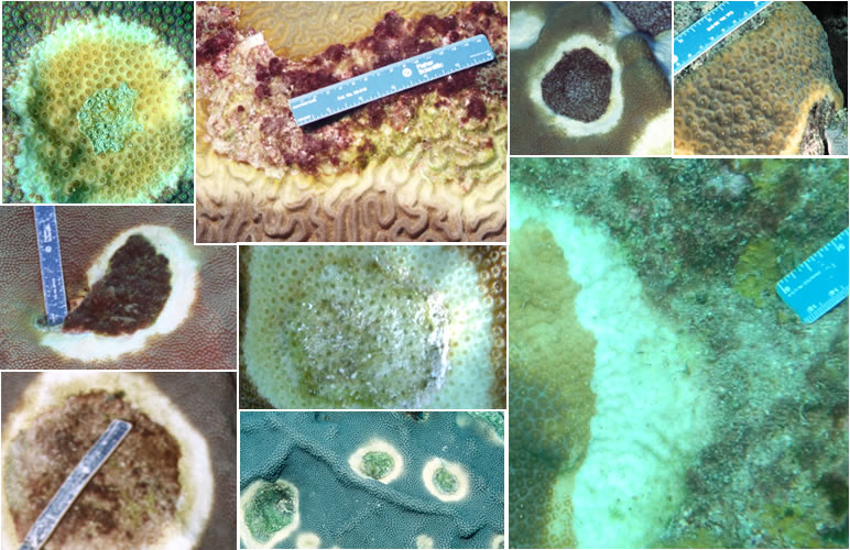
Yellow band disease, YBD (A-I)
Begins as a pale yellow, circular blotch of tissue surrounded by normal tissue (A), or as a narrow band at the edge of a colony (F). These focal (E), multifocal (I) or annular to linear lesions expand in size a few mm to cm per month, coalesce (H) and slowly migrate across the colony (B-C). The leading edge of the band becomes a light pale yellow or lemon color, while tissue first affected gradually darkens prior to dying (A). Small patches (< 1cm) of recent tissue loss may be observed, but skeletal areas are usually colonized by epibionts (G). May be confused with bleaching. Most common in Montastraea annularis (E) and M. faveolata (A,B,C, H, I) also in Diploria strigosa (D), M. cavernosa (F) and other faviids.

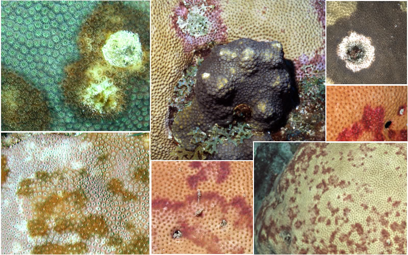
Dark spots disease, DSD (J-P)
Round to irregular, focal to multifocal, dark spots, purple to brown in color, 1-45 cm or more in diameter. Dark spots may grow in size over time, coalesce (J, L), and form irregular to annular bands (M) adjacent to or surrounding exposed skeleton. Affected tissue may be associated with a depression of the coral surface (N) and in some cases spots may appear seasonally. In Siderastrea, purple spots may appear over the surface, or form a band at the edge or within the coral that slowly moves (M, O); Tissue loss is generally very slow. The underlying skeleton often retains the dark pigmentation when exposed. Most common in Siderastrea (K, M, O, P), Montastraea (I, L) and Stephanocoenia (N).
The CDHC is a network of scientists, managers, and agency representatives devoted to understanding coral health and disease.
Funding support provided by NOAA CRCP
Web hosting by NOAA NCCOS
Coral Disease and Health Consortium
Hollings Marine Laboratory
331 Fort Johnson Road
Charleston, SC 29412 USA
Email: cdhc.coral@noaa.gov