Home » Coral Disease » Characterized Diseases » Acropora White Syndromes
Acropora White Syndromes
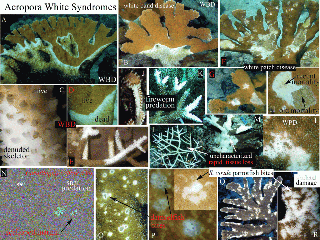
General Diagnostics: Lesions on elkhorn coral (Acropora palmata) and staghorn coral (A. cervicornis) characterized by recent tissue loss separating live tissue and algal-colonized skeleton; no evidence of color change; absence of a pigmented band, no skeletal damage or abnormal growth. Lesions may be focal, multifocal, coalescing, linear or annular. The lesion location, shape and pattern of spread are unique for each syndrome. Snails or fireworms may occur on lesions and they also cause similar patterns of tissue loss.
White Band Disease (WBD) (A-E)
Lesions may be annular or linear, spreading from the base to the tips, or starting at a branch bifurcation and spreading up or down; tissue loss advances 1-100 mm/day. On A. palmata tissue loss may progress up the underside or upper surface of the branch; the band also may encircle the entire branch. Tissue at the lesion margin may be bleached (WBD type II) and small pieces of tissue may be peeling off the skeleton (C) Exposed skeleton is progressively colonized by epibionts.
White Patch Disease (WPD) (F-I)
Also called patchy necrosis, white pox: Lesions are focal to multifocal, coalescing lesions, irregular in shape, completely surrounded by living tissue. Lesions initiate within a branch surface on unaffected tissue (I) or at the margin of an old lesion (H) Colonies may have acute (recent), subacute (colonized) and old lesions (F) lesions radiate out over time and coalesce; resheeting occurs once mortality stops. Lesions have a sharply demarcated leading edge of tissue loss; tissue remnants may be present and corallites may be broken. 1- 80 cm in diameter, Damselfish may be associated with WPD lesions and may cause similar patterns of tissue loss.
Uncharacterized Rapid Tissue Loss (L,M)
White syndromes with diffuse, rapid tissue loss in an irregular pattern that differs from WBD and WPD.
Similar Conditions
Fireworm predation (J-K)
Resembles WBD type II; tissue loss confined to branch tips. Recent lesions lack algal colonization; tissue margin is smooth and not sloughing.
Snail predation (N)
Advances in a linear or annular pattern like WBD, but lesions have a serpiginous scalloped or undulating margin the shape of the snail’s shell; snails are on the lesion or at the colony perimeter.
Damselfish bites (O-P)
Round to irregular, focal, multifocal or coalescing, small (1-3 cm) but expanding lesions often with broken corallites; may form “chimneys”.
Parrotfish predation (Q-R)
Irregular loss of tissue and underlying skeleton often at the edge of the branch; scrape marks and jaw pattern may be visible.
 Official websites use .gov
A .gov website belongs to an official government organization in the United States.
 Secure .gov websites use HTTPS
A lock or https:// means you’ve safely connected to the .gov website. Share sensitive information only on official, secure websites.
