 Official websites use .gov
A .gov website belongs to an official government organization in the United States. Official websites use .gov
A .gov website belongs to an official government organization in the United States. |
 Secure .gov websites use HTTPS
A lock or https:// means you’ve safely connected to the .gov website. Share sensitive information only on official, secure websites. Secure .gov websites use HTTPS
A lock or https:// means you’ve safely connected to the .gov website. Share sensitive information only on official, secure websites. |
Solutions today for reefs tomorrow
Home » Coral Disease » Private: Abiotic Lesions » Predation
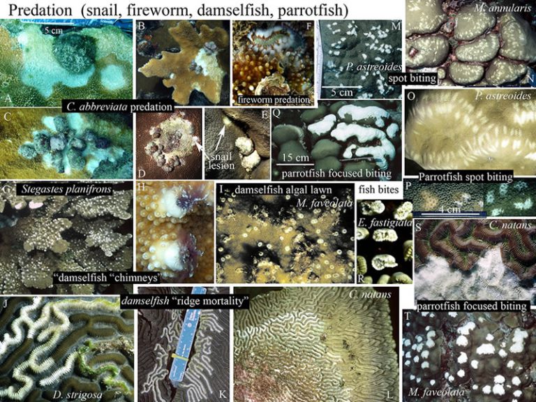
Invertebrate Predation: Check for the presence of corallivores at the lesion margin or the base of the colony.
Hermodice fireworms (F)
Engulf entire branches and protuberances on the colony; uniform and complete removal of tissue from branch tips or projections; smooth, uniform margin between skeleton and live tissue; no evidence of tissue sloughing. Feed mostly at night.
Coralliophila snails (A-E)
Feed in groups (2-70+) at the edge or base of a coral consuming tissue directly under their shell footprint, creating a scalloped pattern. Tissue loss initiating at base or perimeter of coral. The feeding scar progressively radiates out in a linear or annular pattern, but it may extend in an irregular path across the coral. Tissue loss is concentrated on upper surfaces of branches (B) Lesion margin may be irregular, serpiginous, serrated or undulating (A) ; no sloughing of tissue at the interface of exposed skeleton like in WBD, WP and WPD.

Fish Predation: Fish may be seen biting coral; fresh lesions stream mucus. Absence of tissue and skeleton.
Parrotfish predation
Damselfish predation
The CDHC is a network of scientists, managers, and agency representatives devoted to understanding coral health and disease.
Funding support provided by NOAA CRCP
Web hosting by NOAA NCCOS
Coral Disease and Health Consortium
Hollings Marine Laboratory
331 Fort Johnson Road
Charleston, SC 29412 USA
Email: cdhc.coral@noaa.gov