Borger, J. L. (2003). Three scleractinian coral diseases in Dominica, West Indies: Distribution, infection patterns and contribution to coral tissue mortality. Revista de Biologia Tropical, 51(SUPPL. 4).
Borger, J. L. (2005). Scleractinian coral diseases in south Florida: Incidence, species susceptibility, and mortality. Diseases of Aquatic Organisms, 67(3). https://doi.org/10.3354/dao067249
Borger, J. L., & Steiner, S. C. C. (2005). The spatial and temporal dynamics of coral diseases in Dominica, West Indies. Bulletin of Marine Science, 77(1).
Brandt, M. E., & Mcmanus, J. W. (2009). Disease incidence is related to bleaching extent in reef-building corals. Ecology, 90(10). https://doi.org/10.1890/08-0445.1
Cervino, J., Goreau, T. J., Nagelkerken, I., Smith, G. W., & Hayes, R. (2001). Yellow band and dark spot syndromes in Caribbean corals: Distribution, rate of spread, cytology, and effects on abundance and division rate of zooxanthellae. Hydrobiologia, 460. https://doi.org/10.1023/A:1013166617140
Correa, A. M. S., Brandt, M. E., Smith, T. B., Thornhill, D. J., & Baker, A. C. (2009). Symbiodinium associations with diseased and healthy scleractinian corals. Coral Reefs, 28(2). https://doi.org/10.1007/s00338-008-0464-6
Cróquer, A., & Weil, E. (2009). Changes in Caribbean coral disease prevalence after the 2005 bleaching event. Diseases of Aquatic Organisms, 87(1–2). https://doi.org/10.3354/dao02164
Francini-Filho, R. B., Moura, R. L., Thompson, F. L., Reis, R. M., Kaufman, L., Kikuchi, R. K. P., & Leão, Z. M. A. N. (2008). Diseases leading to accelerated decline of reef corals in the largest South Atlantic reef complex (Abrolhos Bank, eastern Brazil). Marine Pollution Bulletin, 56(5). https://doi.org/10.1016/j.marpolbul.2008.02.013
Garzón-Ferreira, J., Gil-Agudelo, D. L., Barrios, L. M., & Zea, S. (2001). Stony coral diseases observed in southwestern Caribbean reefs. Hydrobiologia, 460. https://doi.org/10.1023/A:1013133818360
Gil-Agudelo, D. L., & Garzón-Ferreira, J. (2001). Spatial and seasonal variation of dark spots disease in coral communities of the Santa Marta area (Colombian Caribbean). Bulletin of Marine Science, 69(2).
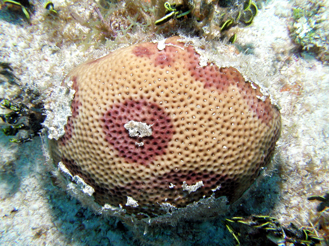
Gil-Agudelo, D. L., Smith, G. W., Garzón-Ferreira, J., Weil, E., & Petersen, D. (2004). Dark Spots Disease and Yellow Band Disease, Two Poorly Known Coral Diseases with High Incidence in Caribbean Reefs. In Coral Health and Disease. https://doi.org/10.1007/978-3-662-06414-6_19
Gochfeld, D. J., Olson, J. B., & Slattery, M. (2006). Colony versus population variation in susceptibility and resistance to dark spot syndrome in the Caribbean coral Siderastrea siderea. Diseases of Aquatic Organisms, 69(1). https://doi.org/10.3354/dao069053
Kaczmarsky, L. T., Draud, M., & Williams, E. H. (2005). Is there a relationship between proximity to sewage effluent and the prevalence of coral disease? Caribbean Journal of Science, 41(1).
Voss, J. D., & Richardson, L. L. (2006). Coral diseases near Lee Stocking Island, Bahamas: Patterns and potential drivers. Diseases of Aquatic Organisms, 69(1). https://doi.org/10.3354/dao069033
Ward, J. R., Rypien, K. L., Bruno, J. F., Harvell, C. D., Jordán-Dahlgren, E., Mullen, K. M., Rodríguez-Martínez, R. E., Sánchez, J., & Smith, G. (2006). Coral diversity and disease in Mexico. Diseases of Aquatic Organisms, 69(1). https://doi.org/10.3354/dao069023
Weil, E., and Croquer, A. (2009) Spatial variability in distribution and prevalence of Caribbean scleracintion coral and octocoral diseases. I. Community-level analysis. Diseases of Aquatic Organisms 83, 195-208.
Weil, E., Smith, G., & Gil-Agudelo, D. L. (2006). Status and progress in coral reef disease research. In Diseases of Aquatic Organisms (Vol. 69, Issue 1). https://doi.org/10.3354/dao069001
Work, T. M., Aeby, G. S., Stanton, F. G., & Fenner, D. (2008). Overgrowth of fungi (endolithic hypermycosis) associated with multifocal to diffuse distinct amorphous dark discoloration of corals in the Indo-Pacific. Coral Reefs, 27(3). https://doi.org/10.1007/s00338-008-0374-7
 An official website of the United States government.
  Official websites use .gov
A .gov website belongs to an official government organization in the United States.
Official websites use .gov
A .gov website belongs to an official government organization in the United States. Secure .gov websites use HTTPS
A lock or https:// means you’ve safely connected to the .gov website. Share sensitive information only on official, secure websites.
Secure .gov websites use HTTPS
A lock or https:// means you’ve safely connected to the .gov website. Share sensitive information only on official, secure websites.